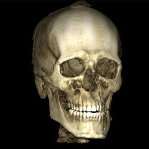
3D Dental X Rays – What Are 3D Dental X Rays
Three-dimensional X-rays capture a true 3-D image of the mouth and allow a dentist to study the mouth in slices,  similar to a CT scan. Three-dimensional X-rays sometimes referred to as a cone beam, capture images of the teeth and mouth. As a result, patients are able to see and understand their own dental X-rays easier. These 3-D X-rays show in great detail many things that 2-D X-rays are not able to capture. Traditional dental X-rays show only two dimensions of a three-dimensional object. This is the case whether the X-rays are exposed on film and held up to the light for evaluation, or taken digitally and viewed on a computer.
Many patients assume when they have an X-ray taken at the dentist and the machine moves around their head that they are getting a 3-D X-ray, but this isn’t true. Very few dentists, less than five percent, have a 3-D X-ray machine. Dr. Anna Smith, owner of Dentistry With TLC in Godfrey, IL was one of the first in the St. Louis region and is the first dentist in the Riverbend area to have a 3-D X-ray machine.
Three-dimensional X-rays make dentistry more predictable and faster for both the patient and the dentist. There are times when a patient’s tooth hurts and a 2-D X-ray shows no problem. However, a 3-D X-ray may reveal an abscess, an infection, or a crack in the root of a tooth. This allows the patient to have their toothache fixed sooner rather than later.
Dr. Smith uses her 3-D X-ray machine before every implant placement. This allows her to see the thickness of the bone and position of the nerves before the surgery. Having the ability to take a 3-D X-ray in her office usually, saves her patients at least one appointment during the implant process. Patients appreciate that she has the 3-D X-ray available in her office because it can make getting an implant three to six months faster than doing the same procedure without a 3-D X-ray.
Three-dimensional X-rays can be taken quickly and comfortably because this type of X-ray does not require anything to be placed inside the mouth. The patient is simply relaxed in a standing, sitting, or wheelchair accessible position and the imaging machine is adjusted to each individual patient. With 3-D X-rays, positioning errors and retakes are a thing of the past, which helps reduce the amount of radiation to each patient.
